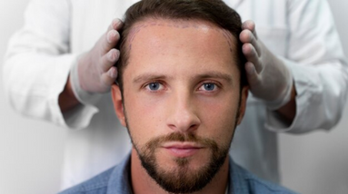
Cavernoma, also known as cavernous malformation or cavernous hemangioma, is a type of vascular abnormality that can occur in the brain or spinal cord. It is characterized by a cluster of abnormally enlarged blood vessels, resembling a “cavern” or small sac.
Cavernomas are typically congenital, meaning they are present at birth, although they can also develop later in life. They are formed by thin-walled blood vessels with irregularly dilated spaces or “caverns.” These blood vessels are prone to leakage and can cause symptoms when they bleed or disrupt nearby brain tissue.
The exact cause of cavernomas is not fully understood, but genetic factors are believed to play a role in their development. Some cases of cavernomas are associated with specific genetic mutations, while others occur sporadically without an identified genetic cause.
Symptoms of cavernomas can vary depending on their size, location, and whether they have caused bleeding or other complications. Some individuals may remain asymptomatic, while others may experience:
The diagnosis of cavernomas is typically made through imaging studies, such as magnetic resonance imaging (MRI) or computed tomography (CT) scans. MRI is particularly useful in visualizing the characteristic appearance of cavernomas.
Treatment options for cavernomas depend on several factors, including the size, location, symptoms, and risk of bleeding or other complications. In some cases, observation with regular imaging studies may be recommended if the cavernoma is small, asymptomatic, and carries a low risk of bleeding.
When treatment is necessary, options may include:
The choice of treatment depends on various factors, and the decision is made on an individual basis, considering the risks and potential benefits for each patient.
Long-term follow-up care is essential for individuals with cavernomas, as they carry the risk of bleeding or other complications. Regular imaging studies and clinical evaluations are necessary to monitor the cavernoma and manage any potential changes or recurrent symptoms.
As we are an independent consulting company we search for the best medical team and establishment for our counselees.
Adress: Abide-I Hürriyet Cad. Merkez Mah. No:110 Kat: 3- Şişli – İstanbul
Phone: +90 545 358 16 69
Mail: info@mkmedicare.com