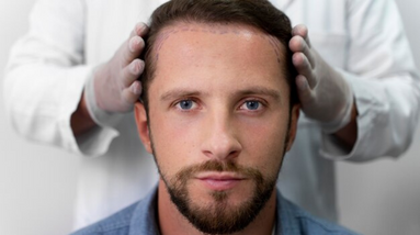
Endoscopic subdural hematoma drainage is a minimally invasive surgical procedure used to remove a subdural hematoma, which is a collection of blood between the dura mater (the outermost membrane covering the brain) and the arachnoid mater (the middle membrane).
Traditionally, subdural hematomas were treated with a craniotomy, which involves making a larger surgical opening in the skull. However, endoscopic techniques have been developed as an alternative, less invasive approach.
During endoscopic subdural hematoma drainage, the surgeon makes a small incision in the scalp and creates a small hole in the skull (burr hole). An endoscope, which is a thin tube with a camera and light source, is inserted through the burr hole and guided to the site of the hematoma.
The endoscope provides visual guidance to the surgeon, allowing for direct visualization of the hematoma and surrounding structures. Specialized instruments are used to evacuate the clot and remove any trapped blood or debris.
Endoscopic subdural hematoma drainage offers several potential benefits compared to traditional craniotomy, including:
However, not all subdural hematomas are suitable for endoscopic drainage. The decision to use this technique depends on several factors, including the size and location of the hematoma, the patient’s overall health, and the surgeon’s expertise and preference.
As with any surgical procedure, there are risks associated with endoscopic subdural hematoma drainage, such as infection, bleeding, damage to surrounding structures, or recurrence of the hematoma. It’s important to consult with a qualified neurosurgeon who can assess the individual case and determine the most appropriate treatment approach.
After the procedure, patients typically require monitoring in the hospital for a period of time to ensure proper recovery and to address any potential complications. Postoperative care may include pain management, wound care, and close follow-up with the medical team.
As we are an independent consulting company we search for the best medical team and establishment for our counselees.
Adress: Abide-I Hürriyet Cad. Merkez Mah. No:110 Kat: 3- Şişli – İstanbul
Phone: +90 545 358 16 69
Mail: info@mkmedicare.com