
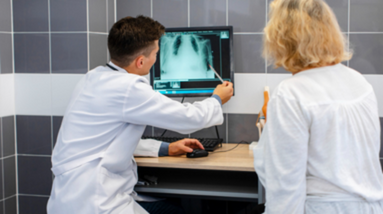
Chest X-ray is a type of radiological imaging test that produces images of the chest, including the heart, lungs, and other organs in the thoracic cavity. The chest X-ray is commonly used to evaluate respiratory symptoms such as cough, shortness of breath, chest pain, or to assess injuries to the chest. It is also used as a screening tool to detect various medical conditions, including pneumonia, tuberculosis, lung cancer, and heart failure. The procedure is non-invasive, quick, and painless. The patient stands in front of the X-ray machine and holds their breath for a few seconds while the X-ray is taken. The radiation dose is relatively low and considered safe. Chest X-rays can be interpreted by radiologists or other healthcare providers who are trained to read X-ray images.
As we are an independent consulting company we search for the best medical team and establishment for our counselees.
Adress: Abide-I Hürriyet Cad. Merkez Mah. No:110 Kat: 3- Şişli – İstanbul
Phone: +90 545 358 16 69
Mail: info@mkmedicare.com